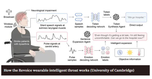
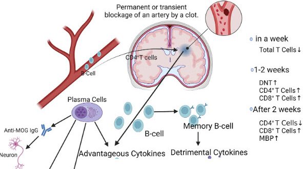
Your Stroke / Brain Injury Recovery Starts Here


ARNI home-based training and guidance for your rehab is POWERFUL. Accept no substitute.

2026 marks 25 years of ARNI: Click Here to Claim your £50 off Full Set of Stroke Rehab 7 DVDs TODAY!

News

Not a News Item today, but an important Technical Reminder: for our stroke survivors and their professional therapists and ARNI neuro-instructors, navigating the complexities of recovery, understanding the mechanics of symptoms is critical. One of the questions that these groups ask experts in stroke is exactly why spasticity is negatively affected by movement speed.

So, one of the most defining characteristics of spasticity after stroke is its reliance on velocity – in effect, the question isn’t just if there is resistance, but how quickly you move. This phenomenon is rooted entirely in the intricate neurophysiology of the spinal cord reflex arc and the disruption caused by an upper motor neuron (UMN) lesion.

In a healthy system, our muscle tone is finely tuned by a balance of excitatory and inhibitory signals. After a UMN lesion, perhaps from a stroke or spinal cord injury, this descending inhibitory control is diminished or lost. The spinal circuitry becomes disinhibited, hypersensitive to input. The key sensory players here are the muscle spindles; specialised receptors within the muscle belly that detect changes in length and the speed of those changes.

The primary afferent fibers (Group Ia afferents) innervating these spindles are exquisitely sensitive to the velocity of a stretch. When a therapist or trainer moves a limb which is in in a position of spasticity, stretching the muscle, these velocity-sensitive fibers fire robustly. The faster the movement, the greater the electrical signal transmitted back to the spinal cord. In the disinhibited state, this heightened afferent barrage leads to an exaggerated excitation of the alpha motor neurons, the final common pathway for muscle contraction.

The result is an abnormally strong, brisk, exaggerated involuntary efferent contraction, which manifests clinically as increased resistance proportional to the speed of the stretch applied. This mechanism distinguishes spasticity from other forms of hypertonia like rigidity, which is velocity-independent. Understanding this principle is crucial for effective therapy; it’s why slow, sustained stretches are often more effective than quick movements in managing spasticity and why careful grading of speed is vital during functional tasks…

So, essentially, speed is the key trigger! The quicker the movement, the stronger the sensory feedback that bypasses normal modulation, resulting in that characteristic resistance we measure clinically, using scales like the Modified Ashworth Scale (MAS) and the Tardieu Scale (evaluating stretch reflex at different speeds).

For more about spasticity, please see this post on the ARNI site from 2019: https://arni.uk.com/spasticity-after-stroke-will-my-hand…/

If you’ve had a stroke and like engaging with nature, Izzy and Rocksy, two friends of ARNI Stroke Rehab UK (Occupational Therapy students from the University of Southampton), would love to hear from you as they’re doing a study on how people with neurological conditions like stroke experience and engage with nature-based activities, such as gardening, outdoor walks, training in nature, conservation projects etc… please help them!

Nature-based rehabilitation (NBR) delivered as part of neurorehabilitation is a brilliant and developing approach.

To help the Southampton University students do research and create a protocol, do read on and consider doing a quick, helpful computer-based interview (no need to go anywhere) with them if you can spare the time!

The Southampton students would like to understand better, things like:

  • What does nature mean to you?
  • What helps or prevents you to engage with it?
  • How does it support or challenge your wellbeing and daily life?

As you know, each year in the UK, around 100,000 people experience a stroke contributing to a community of approximately 1.3 million stroke survivors, more than half of whom live with a disability.

Post-stroke fatigue is one of the most common and challenging symptoms to manage. Dong et al, (2025), reported that 45.8 % of individuals in their study experienced post-stroke fatigue, highlighting the need for interventions that address depression, improve quality of life and restore activities of daily living.

Living with a neurological condition, such as stroke, involves navigating a complex mix of physical, cognitive and emotional challenges that affect everyday life, wellbeing and connection with others. Many people experience fatigue, anxiety, reduced confidence or difficulty taking part in activities that once felt meaningful.

As interest grows in holistic, person-centred approaches to rehabilitation, nature engagement is emerging as a promising way to support wellbeing for individuals living with long-term conditions.

Nature engagement is endless and broad, and it can range from a forest walk, gardening, horticultural engagement, horse riding and many more possibilities!

Research suggests that spending time in natural environments can offer meaningful benefits such as improvements in mood, confidence, social connection, and overall quality of life for people with neurological conditions. Many people describe nature as a space where they feel more motivated, more independent and more able to challenge themselves in their rehabilitation journey.

To date, despite all these findings, there remains limited research exploring the personal experiences of nature engagement for people with neurological conditions.

Izzy and Rocksy want to contribute to stroke survivors – they state that ‘understanding these perspectives is essential for developing accessible, effective and meaningful nature-based programmes that actually reflect what stroke survivors value and need’.


This study aims to gather the experiences of people who have experienced stroke and who take part in any form of nature-based activity.

The Southampton students would like to understand better, things like:

  • What nature means to you?
  • What helps or prevents you to engage with it?
  • How does it support or challenge your wellbeing and daily life?

Your insights can help shape future stroke rehabilitation approaches and ensure that nature-based programmes are designed with lived experience at their core.

Your insights will help us understand what makes it easier or harder to take part in these kinds of activities and how they can be made more accessible for everyone.

What will happen to me if I take part?

  • If you agree to take part, we will ask you to fill in a consent form and a short form to provide background information about you.
  • You will be invited to a one-to-one interview lasting around 45–60 minutes. This will take place online via Microsoft Teams at a time that suits you.
  • During the interview, we will ask you about your experiences with nature-based activities, the factors that make participation easier or harder, and what you feel the benefits are.
  • The interview will be video recorded on Microsoft teams (with your consent) to help us accurately transcribe what is said. The recording will be securely deleted once the transcription is complete. We will remove any identifying information about you from the transcript.
  • This transcript will be securely stored on a University of Southampton SharePoint site, accessible to the research team. You do not need to answer any questions that make you feel uncomfortable. You can take breaks or stop the interview at any time..

Are there any benefits in my taking part?

Yes! Your views will contribute to raising awareness and improving understanding of how nature-based interventions are experienced and delivered.

The Southampton students hope that your participation will help inform future practice and promote greater inclusivity for people living with long term neurological conditions. Please contact them on either im2g23@soton.ac.uk or ra11g23@soton.ac.uk

The findings from this study will also be used to teach undergraduate students about the value and application of nature based programmes.

The aim is to gain insight into barriers and enablers that impact engagement in nature-based programmes, and to understand how these activities can be improved and sustained to better support the wellbeing of people with long-term neurological conditions.

The project is not funded in any way.

* Researchers: Izzy Mason, Undergraduate Researcher (BSc Occupational Therapy) University of Southampton and Rocksy Antonygnaneswaran, Undergraduate Researcher (BSc Occupational Therapy), University of Southampton.

* Supervisor: Long-time friend of ARNI Stroke Rehab UK Charity: Dr Leisle Ezekiel (lezekiel@soton.ac.uk)


Dong, Y., Tang, L., Salwismawati Badrin, Salziyan Badrin and Wu, J. (2025). Factors associated with post-stroke fatigue among stroke survivors: a cross-sectional study. PeerJ, 13, pp.e19052–e19052.

In the high-stakes world of stroke medicine, the mantra is simple and urgent: ‘Time is brain’. Every minute that a major blood clot blocks the flow to the brain, more vital tissue dies. Doctors race against the clock to diagnose, locate the blockage, and intervene. But what if we could give doctors an extra hour in that critical race? A groundbreaking new study suggests we can do exactly that, all thanks to an innovative artificial intelligence imaging tool.

Researchers have found that this sophisticated AI can help doctors spot deadly large vessel occlusions (LVOs);the most severe and debilitating type of stroke-causing clots, an average of more than an hour earlier than current clinical workflows allow.

The AI doesn’t replace the expert eye of a radiologist or neurologist; instead, it acts as an incredibly efficient triage system and second set of eyes. It rapidly processes complex brain scans and immediately flags critical findings, pushing relevant cases to the top of the queue for human review. This efficiency drastically cuts down the time from when a patient arrives at the emergency department to when the critical treatment decision is made…

Earlier diagnosis means faster initiation of life-saving treatments like mechanical thrombectomy (physically removing the clot). The difference between intervention at two hours versus three hours can mean the difference between a patient making a full functional recovery or suffering a severe, lifelong disability.

In our fast-paced world, stress is often dismissed as just ‘part of life’. We talk about it in passing, joke about our caffeine intake, and rarely view it as a serious health threat. However, emerging research is strengthening the vital connection between chronic, psychosocial stress and a significantly increased risk of stroke. This invisible emotional burden may be quietly damaging  brain.

Multiple extensive studies and large-scale meta-analyses published last month (Frontiers Neurol. 2025 Nov 6;16:1669925) , encompassing nearly a million (950,000) participants, confirm a moderate yet significant association between self-perceived chronic stress and stroke risk. The data is compelling, suggesting that stress impacts our health with a magnitude similar to factors like diabetes, making it a critical, and often overlooked, modifiable risk factor.

So, how does the pressure of a job or relationship translate into a physical blockage or bleed in the brain? The connection lies in the body’s ‘fight-or-flight’ response. When stressed, our systems flood with hormones like cortisol and adrenaline. While useful for escaping immediate danger, constant elevation of these hormones over years leads to sustained high blood pressure, chronic inflammation throughout the cardiovascular system, and actual damage to artery walls. This creates a perfect environment for clots to form and blood vessels to harden (atherosclerosis).

Furthermore, chronic stress often pushes people toward unhealthy coping mechanisms. Poor sleep quality, emotional eating, increased smoking, lack of physical exercise and higher alcohol consumption become common responses. These lifestyle choices independently pile on further risk factors for stroke, creating a vicious cycle of physical and emotional strain.

Intriguingly, recent research published in the journal Neurology® points to a potential gender disparity. One specific study focusing on young adults indicated that women reporting moderate to high stress levels faced a notably higher risk of stroke compared to men with similar stress burdens. This highlights a need for targeted awareness and stress management strategies tailored to different demographics.

The main takeaway is clear: managing stress is not just about mental well-being or ‘feeling better’… it’s a fundamental component of preventative medicine and physical health. Incorporating simple, consistent practices like mindfulness, regular walks, finding time for hobbies or seeking professional support can make a tangible, life-saving difference.

Frontiers Neurol. 2025 Nov 6;16:1669925 https://pmc.ncbi.nlm.nih.gov/articles/PMC12631615/

One of the biggest challenges for survivors is consistently performing the intensive physical therapy needed to regain movement.. especially for those dealing with hemiparesis, simple upper-limb exercises can feel impossible or deeply discouraging. Enter a brilliant innovation that’s changing the game: the Powered Rehab Skateboard, developed by Prof. Kenneth Fong and team at Hong Kong Polytechnic University.

The device is a sophisticated, motorised platform engineered specifically for the more-affected upper limb: it gently supports the arm and guides it through precise, repetitive therapeutic motions.

The core of the device is a wheeled, end-effector-based robotic system (a ‘skateboard’ on a table) which supports the patient’s arm and guides it through specific movement patterns (e.g., left-right, forward-backward, circular, and figure-eight motions)

It features an integrated torque sensor that detects the user’s active force in real-time. This allows the device to measure how much effort the patient is putting into the exercise. It also  uses a control system that interprets the sensor data to adjust the level of assistance dynamically. This system offers multiple operational modes: passive, assistive and resistive.

A machine learning algorithm (in advanced versions of the related research) captures activity data and adapts the therapy in real-time, ensuring optimal challenge and support for the individual’s specific stage of recovery. For home use, the device includes a micro edge detection sensor that triggers an alarm and stops operation if it approaches the edge of a table, preventing accidents.

In essence, the technology provides a ‘smart’ physical therapist at home, ensuring the patient receives the correct intensity and type of repetitive exercise necessary for neurorehabilitation and motor learning. 

It’s primarily an academic and clinical research innovation at this stage, so not yet a commercial product you can simply buy from a medical supply store today. To find out when it might become commercially available for UK stroke survivors, the best course of action is to follow updates here at ARNI – we’ll find out and let you know when we hear official announcements from Prof. Kenneth Fong’s research team. While we can’t buy it off the shelf yet, the promise it holds for home-based recovery is undeniable.

Exciting news is emerging from the latest clinical research; the innovative SuperNOVA Stent (Neurovascular Ostial Advanced Stent Retriever) has just been demonstrated to be both safe and effective for treating acute ischemic strokes in clinical trials. This is a significant development in the world of endovascular therapy, offering new possibilities for improving outcomes for survivors.

For those unfamiliar, mechanical thrombectomy involves physically removing a blood clot from a blocked artery in the brain using specialised devices delivered via a catheter. This procedure has revolutionized severe stroke care since its proven efficacy a decade ago. The SuperNOVA stent is a next-generation device designed to potentially offer improved performance in retrieving those tricky, large vessel occlusions, aiming for faster and more complete restoration of blood flow to the brain. The recent studies have confirmed its efficacy, showing high rates of successful recanalisation with excellent safety profiles for patients involved in the trials.

This breakthrough means clinicians may soon have another highly effective tool in their arsenal to combat strokes rapidly. Faster, more complete clot removal directly translates to better outcomes, reduced long-term disability and improved quality of life for survivors. Regarding its introduction for clinical practice in the UK, the path forward involves regulatory approval and NHS adoption processes. Following successful clinical trials, the device must receive the appropriate regulatory certification (such as a UKCA mark, previously CE mark, depending on current regulations) confirming it meets safety and performance standards.

Once certified, NHS trusts and National Institute for Health and Care Excellence (NICE) will need to evaluate the cost-effectiveness and clinical benefits compared to existing stent retrievers. This process can take time, often several months to a couple of years depending on the complexity and priority. So, while a promising development, ARNI Stroke Rehab UK comments that immediate availability in every UK hospital this spring is unlikely! Clinicians and patients can expect to see it introduced into specialist neurovascular centres gradually over the next one to three years, subject to successful regulatory hurdles and local commissioning decisions.

Question for you? What does 3-D printing and predicting if you’re going to have a stroke got to do with each other? Can you guess?

Well – here’s ARNI Stroke Rehab UK’s prediction of the very near future in a hospital near you: you go to have a CT scan, a technician rapidly 3-D prints your blood vessel model, tests your blood response and then uses AI to predict your stroke risk years in advance.

Why do we reckon this? Because researchers at the University of Sydney have just developed a groundbreaking new 3D printing technique that can generate anatomically accurate replicas of blood vessels in a mere two hours. That’s down from 10 hours using older methods! This device represents a significant leap forward in biomedical engineering.

The challenge of understanding and treating stroke is fundamentally tied to the incredibly complex architecture of the human vascular system. Replicating the intricate network of blood vessels to study how clots form and behave has long been a time-consuming and often imprecise process, frequently relying on animal models which don’t perfectly mimic human anatomy.

By utilising the specific CT scans of actual stroke patients, the researchers can create incredibly realistic models that mimic the exact fluid dynamics and anatomical quirks of a real person’s vascular system. These ‘vessel twins’ are not just static models; they are functional and allow scientists to literally watch a blood clot form under a microscope in a highly realistic environment.

The implications of this speed and accuracy are vast for stroke research. The models are already being used to study the exact mechanisms of blood clot formation, helping researchers understand why some individuals are more prone to certain types of strokes. More importantly, this technology provides an ethical and efficient alternative to traditional methods. Researchers can now trial new clot-busting drugs, test different surgical devices and experiment with medicine approaches on these replicas without relying on animal testing. This accelerates the research timeline, reduces costs and provides more accurate data relevant to human patients.

When we think about stroke, our minds immediately go to the brain: damage, recovery, rehabilitation. But the reality is that a stroke is a whole-body event, triggering complex physiological changes far beyond the site of the injury. For many stroke survivors, this manifests in a surprising and often frustrating way: gastro-intestinal problems. Now, thanks to groundbreaking research from scientists at The University of Manchester, we are beginning to understand why this happens and what we might be able to do about it.

The research, just published in Brain, Behaviour and Immunity, adds to the emerging idea of the ‘gut-brain axis’ by shedding new light on the intricate connection between the brain and the gut, specifically focusing on the gut’s immune system. What the Manchester team discovered is that a stroke doesn’t just cause immune suppression; it actively triggers a disruption in the delicate balance of immune responses within the digestive tract. Think of your gut lining as a highly secure barrier; after a stroke, this barrier can become ‘leaky’ and inflamed due to these immune system changes.

This ‘leaky gut’ and the ensuing inflammation are what contribute directly to the various gastro-intestinal issues that survivors often face, from digestive discomfort and motility issues to a heightened risk of infection. It’s a vicious cycle where a brain event affects the gut, and the gut inflammation can, in turn, impede overall recovery.

In the past, these gastrointestinal issues were often treated symptomatically without fully grasping the root cause linked to the initial neurological event. Now that scientists have a clearer picture of the immune system’s role, it opens the door to new therapeutic strategies; by specifically targeting these immune changes in the gut, doctors might be able to prevent these secondary health complications from occurring in the first place, or manage them much more effectively.

This innovative research seems to underscore the importance of a holistic approach to stroke recovery, focusing not just on the brain and limbs but on overall systemic health.

Our NHS works incredibly hard, but logistical challenges, particularly in getting patients to specialist centres for immediate CT or MRI scans, can sometimes lead to crucial delays. This is why the development of AI-Stroke, a revolutionary initiative aiming to turn any smartphone into a rapid stroke-assessment tool, shows promise for future UK stroke survivors and their families.

AI-Stroke’s core function is simple yet impactful: a paramedic in an ambulance, a triage nurse in a busy A&E, or perhaps eventually even a trained professional in a remote GP surgery, can record a 30-second video of a person suspected of having a stroke. The sophisticated AI then immediately analyses that footage. It scrutinises key indicators of stroke signs that align with the vital F.A.S.T. test: facial symmetry and within seconds, provides an alert and assessment score.

By providing an instant, reliable diagnostic aid right at the bedside or in the ambulance, AI-Stroke could drastically cut down the time from symptom onset to appropriate treatment. It allows for immediate communication with specialist stroke teams, preparing them for the patient’s arrival and potentially allowing for faster decision-making regarding life-saving interventions like clot-busting medication or mechanical thrombectomy.

This innovation, which today announced the completion of a US$4.6 million seed round, could drastically reduce diagnosis time and bring expert assessment capabilities to ambulances, rural areas or busy ERs, bringing expert-level diagnostic support into the hands of frontline healthcare workers everywhere. By shortening diagnostic pathways and ensuring the right patients get to the right specialist centre faster, AI-Stroke could be a genuine lifesaver for countless future UK stroke survivors.

Post-stroke pain is a debilitating reality for a significant number of stroke survivors. Whether it manifests as central post-stroke pain (a chronic, often severe neuropathic pain) other musculoskeletal issues, managing it effectively without impairing movement or cognitive function has been a persistent clinical challenge.

The reliance on broad-acting pain medications often comes with undesirable side effects like drowsiness or motor impairment. However, a recent and highly promising neuroscientific discovery offers a novel and potentially game-changing solution. Researchers have identified a specific mechanism for pain activation after stroke, which could lead to far more targeted and effective relief.

The key to this discovery lies in a specific enzyme that neurons release from outside the cell: the Vertebrate Lonesome Kinase, or VLK. This enzyme was found to be a critical player in activating pain signalling pathways that are often heightened after a neurological event like a stroke. The mechanism is intricate but the implications are straightforward: VLK essentially acts as a switch, turning on or ramping up the pain signals that contribute to chronic discomfort.

The truly exciting aspect of this finding is the specificity of the VLK enzyme’s role. In preclinical studies, researchers demonstrated that by removing or blocking this particular enzyme, they could effectively dull post-stroke pain in the affected areas

Crucially, and what sets this potential therapy apart from many existing pain management options, this reduction in pain did not come at the cost of normal motor function or sensation. The ability to manage pain without broad systemic side effects is a massive leap forward in improving the quality of life for stroke survivors.

This research points toward a future where pain management can be highly targeted, focusing precisely on the mechanisms of stroke-induced pain without the trade-offs often associated with current medications. It opens the door for a new class of analgesics that work on blocking the VLK pathway. While this is still a foundational scientific discovery and not yet a developed treatment, it provides immense hope that more effective and less impairing pain relief is on the horizon for the millions of people worldwide living with chronic post-stroke pain…


We are on Facebook

ARNI