For many stroke survivors and their families, the transition back to independent living is often overshadowed by the persistent fear of falls. This anxiety is well-founded, as balance issues and hemiparesis can significantly increase the risk of accidents in the home. However, advancements in smart home technology are providing new layers of security, most notably through the introduction of the Kami Fall Detect Camera. This innovative device represents a significant leap forward from traditional ‘pendant’ alarms, offering a non-intrusive way to ensure help is always at hand without the need for the survivor to wear a physical device or manually trigger an alert during a crisis.
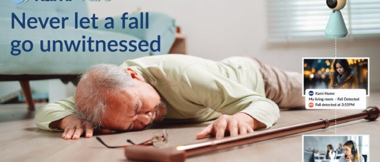
The Kami Fall Detect Camera is a specialised smart security device that utilises advanced Edge AI to monitor for falls in real time. Unlike standard security cameras that simply record movement, this system is specifically programmed to recognize the distinct human posture and motion associated with a fall. When a fall is detected, the camera immediately sends an automated alert to designated family members or caregivers via a smartphone app. This instant notification is crucial for stroke survivors who may experience a ‘long lie’ after a fall; a dangerous period where they are unable to get up or call for help, which can lead to further medical complications like dehydration or pressure sores.
One of the most significant benefits for stroke survivors is the privacy-conscious design of the technology. The camera uses AI to process information locally on the device rather than in the cloud, and it can be configured to represent people as blurred shapes or ‘stick figures’ to maintain dignity while still providing clear evidence of an emergency. Furthermore, the system features two-way audio, allowing caregivers to speak directly to the survivor immediately after a fall is detected, providing vital reassurance while help is on the way. This connection can be a lifeline for someone who may be disoriented or unable to move following a fall.
In terms of investment, the Kami Fall Detect Camera is designed to be an affordable alternative to expensive monthly monitoring services. In the UK, the device typically retails for approximately ninety-nine pounds, though prices can vary slightly depending on current promotions. While there is an optional cloud subscription for extended video history, the essential fall detection and instant alerting features are designed to provide immediate value out of the box. This makes it a cost-effective addition to a home rehabilitation environment where safety is the top priority.
For those looking to purchase the device within the UK, it is most readily available through major online retailers. The official YI Technology and Kami Home stores on Amazon UK are the primary hubs for purchasing, ensuring that customers receive the latest version of the hardware with local warranty support. It can also be found through specialised smart home and independent electronics retailers online. By integrating this kind of intelligent monitoring into the home, stroke survivors can enjoy a greater sense of autonomy… and their loved ones can find peace of mind knowing that technology is keeping a watchful, protective eye on their recovery journey.