Your Stroke / Brain Injury Recovery Starts Here


ARNI home-based training and guidance for your rehab is POWERFUL. Accept no substitute.

2026 marks 25 years of ARNI: Click Here to Claim your £50 off Full Set of Stroke Rehab 7 DVDs TODAY!

News

A 2025 scoping review titled ‘Stroke at the Hairdresser’s Chair’, which was published in the American Journal of Emergency Medicine, synthesised data from 22 previous studies over five decades to provide a comprehensive look at Beauty Parlor Stroke Syndrome (BPSS), a rare but clinically significant phenomenon.

The review analysed 54 reported cases, finding that a significant majority (79.63%) were women, with occurrences linked to neck hyperextension during salon visits, dental procedures, and other activities. Researchers highlighted that while BPSS is uncommon, its symptoms can overlap with those of a classic stroke, emphasising the importance of thorough patient history and neuroimaging for an accurate and timely diagnosis. The study concluded that preventative strategies, such as providing neck support in salons and raising awareness among both clients and professionals, are essential for mitigating this risk. 

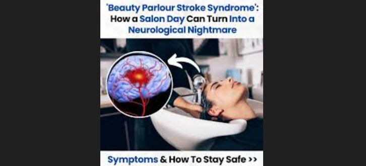
The term ‘Beauty Parlor Stroke Syndrome’, while it may seem colloquial, points to a clinically significant phenomenon: vertebral artery dissection or compression caused by sustained hyperextension of the neck. The setting of a salon shampoo bowl is the most commonly cited trigger, where a patient’s head is tilted backward over a basin’s edge for an extended period, creating mechanical stress on the vessels supplying the brain’s posterior circulation. First described by Dr. Michael Weintraub in 1993, the syndrome highlights how seemingly innocuous, everyday activities can precipitate serious neurological injury, particularly in susceptible individuals.

The underlying pathophysiology centres on compromise of the vertebral arteries, which ascend through the cervical spine to join and form the basilar artery. This critical system supplies blood to the brainstem, cerebellum, and posterior cerebrum. When the neck is hyperextended, especially against a hard surface, this can cause a vessel wall to dissect; a tear that can lead to intramural hematoma formation, luminal occlusion or the release of a clot that embolises to the brain. In other cases, external compression from osteophytes or soft tissues can temporarily restrict blood flow, a phenomenon known as vertebrobasilar insufficiency. The resulting posterior circulation strokes can manifest with symptoms such as dizziness, vertigo, ataxia, visual disturbances, and slurred speech, with symptom onset often delayed for hours or even days.

Epidemiological data, predominantly from case reports and small case series, indicates that while the syndrome is rare, it is disproportionately reported in women. For example, a recent scoping review identified that 79.63% of the 54 cases reviewed were female, with a wide age range from teenagers to the elderly. These findings are critical, as they underscore the need for awareness among patients and salon professionals alike. Risk factors for cervical artery dissection extend beyond the salon, encompassing other activities involving neck hyperextension, such as yoga, painting ceilings, or sudden movements like sneezing. Individuals with pre-existing vascular conditions, such as atherosclerosis, fibromuscular dysplasia or connective tissue disorders, may be at a heightened risk, though the syndrome can also affect young, otherwise healthy individuals.

Raising awareness in the beauty and wellness industries is a crucial preventative measure. Salons can mitigate risk by providing proper neck support, such as rolled towels or cushions, and adjusting chairs to reduce the degree of cervical hyperextension. Patients should be encouraged to communicate any discomfort or neck pain immediately during a shampoo. While the overall risk remains low, the potential for devastating long-term disability; from permanent balance issues to severe neurological deficits… warrants increased vigilance. For clinicians, maintaining a high index of suspicion is essential, particularly when evaluating patients presenting with posterior circulation symptoms following recent neck manipulation. A comprehensive patient history that includes recent activities is vital for accurate and timely diagnosis through neuroimaging. Ultimately, the phenomenon of ‘Beauty Parlour Stroke Syndrome’ is not an urban legend but a clinically confirmed, though rare, condition that necessitates proactive awareness and preventative strategies.

The journey of regaining mobility after a stroke can be arduous, often characterised by the persistence of gait asymmetries and diminished walking endurance. Traditional robotic exoskeletons have offered a partial solution, providing mechanical assistance in structured rehabilitation settings.

However, the inherent variability in stroke-affected gait patterns presents a significant challenge, as many devices struggle to adapt effectively to the unique and often unpredictable movements of individual patients. This rigid approach can inadvertently impede natural joint movements and potentially lead to user discomfort and fatigue, undermining the therapeutic intent.

A groundbreaking development emerges from the George W. Woodruff School of Mechanical Engineering at Georgia Tech, where Associate Professor Aaron Young and his research team have pioneered an AI-powered exoskeleton designed to assist lower limb mobility for stroke survivors. Their innovation addresses the limitations of conventional exoskeletons by introducing a system that prioritises user adaptability and intuitive control. The core of this advancement lies in its AI-driven control architecture which is capable of learning and adapting to the user’s specific gait patterns in real-time.

The team’s research, just published in IEEE Transactions on Robotics, focuses on developing a neural network that functions akin to the human brain… processing sensory inputs from the exoskeleton to precisely modulate assistance for each step.

This intelligent system learns a patient’s unique gait within a remarkable timeframe of just one to two minutes, significantly reducing the laborious manual tuning traditionally required by engineers. The AI continuously monitors and adjusts its support, ensuring the exoskeleton remains synchronised even as the patient’s stride shifts or improves. This results in a 70% reduction in errors compared to standard exoskeletons in tracking stroke patients’ walking patterns, marking a substantial leap in precision and responsiveness.

Beyond the immediate benefits observed in the lab, such as reducing the metabolic cost of walking and improving gait efficiency, this technology holds broader implications. The same adaptive approach could prove invaluable for older adults experiencing age-related muscle weakness, individuals with conditions like Parkinson’s or osteoarthritis, and children with neurological disabilities.

Furthermore, the team has addressed the challenge of data interoperability across different exoskeleton systems by developing software that functions as a universal adapter, allowing the AI to integrate with various devices with minimal calibration. This eliminates the need for extensive retraining when switching between exoskeletons, further streamlining the rehabilitation process.

Clinical trials are now underway to rigorously evaluate the exoskeleton’s performance across a wide range of daily activities.

Stroke remains a leading cause of long-term disability and a major global health burden, with current therapeutic options for acute ischemic stroke limited by a narrow therapeutic window and the formidable challenge of the blood-brain barrier (BBB). Conventional drugs often fail to penetrate the BBB effectively, resulting in poor drug bioavailability in the brain and systemic side effects.

But recent, significant preclinical and early trial data have demonstrated the potential of nanotechnology for advancing stroke treatment by overcoming key limitations of conventional therapies. Nanoparticles, designed to carry therapeutic agents across the formidable blood-brain barrier (BBB), have shown promise in delivering neuroprotective, anti-inflammatory, and thrombolytic drugs directly to ischemic brain tissue.

By encapsulating drugs, these nanocarriers can enhance stability, increase bioavailability, extend circulation time, and enable targeted delivery, thereby reducing the necessary dosage and minimizing off-target side effects. Biomimetic nanoparticles, derived from cell membranes such as platelets or neutrophils, have exhibited superior biocompatibility and targeted delivery to ischemic lesions in animal models by leveraging natural cellular homing mechanisms.

However, the successful clinical translation of this technology faces significant challenges. Further advancements are needed to improve delivery efficiency and simplify complex formulations to ensure predictable and consistent performance. Issues such as cost-effective large-scale production, inconsistent targeting, long-term stability and potential toxicity of the nanoparticles themselves must be addressed.

Bridging the gap between promising preclinical results and effective clinical therapies for future successes, requires concerted research into optimising formulations and navigating complex regulatory frameworks needed for  safe and reproducible clinical translation.

In the high-stakes world of acute stroke care, where every second counts, the speed and efficacy of intervention are paramount to saving brain cells and minimising long-term disability. A revolutionary breakthrough from Stanford University’s Department of Mechanical Engineering and Neuroimaging, published in Nature in June 2025, promises dramatic possibilities. Researchers, including Renee Shao and Jeremy Heit, have unveiled a novel spinning micro-device, the ‘milli-spinner,’ designed to remove brain clots with unprecedented precision and effectiveness.

Current thrombectomy procedures for large vessel occlusion (LVO) strokes, while effective, still have significant limitations. These methods often involve either aspiration catheters that can struggle with larger clots or stent retrievers that grapple and pull the clot, risking fragmentation and dispersal of pieces into smaller, more difficult-to-reach vessels. This can lead to incomplete recanalisation and further brain damage.

The milli-spinner, by contrast, operates on a fundamentally different and more elegant principle. As a tiny, catheter-delivered tool, it utilises a combination of localised suction, compression, and shear forces generated by its rapid spinning action to gently and efficiently process the clot.

The device works by first applying localised suction to secure the clot against the tip of the catheter. The subsequent rapid rotation then creates shear forces that cause the fibrous protein mesh of the clot (fibrin) to roll into a tight, compact ball, shrinking its volume significantly—by up to 95% in preclinical tests.

This ‘fibrin-balling’ action effectively expels the trapped red blood cells, which can then safely continue flowing, leaving behind a manageable, dense clot that is easily removed via suction. This innovative mechanism drastically reduces the risk of fragmentation and subsequent distal embolisation, which are common and serious complications of existing methods.

The results from preclinical studies using flow models and animal subjects were nothing short of remarkable. In tests on tough, fibrin-rich clots that are notoriously difficult to treat with existing devices, the milli-spinner achieved a 90% first-pass success rate in restoring blood flow, a significant leap from the 11% success rate of conventional tools for these cases.

Overall, the device more than doubled the efficacy of current technology, suggesting a major paradigm shift in interventional neurosurgery. Jeremy Heit, Chief of Neuroimaging and Neurointervention at Stanford, called the milli-spinner ‘a sea-change technology,’ projecting that it could save tens of thousands of lives and substantially reduce disability if translated successfully to clinical practice.

The potential impact of this technology extends beyond just higher survival rates. The minimally invasive and highly precise nature of the milli-spinner suggests it could also lead to better functional outcomes and a reduced risk of long-term disability, a critical factor for stroke patients and their families. By achieving faster and more complete clot removal, the device maximises the salvageable brain tissue, a key determinant of post-stroke recovery.

While the device is still undergoing further development and requires clinical trials for human use, a company has already been launched to license the technology from Stanford and accelerate its path to market. The milli-spinner represents a brilliant fusion of engineering innovation and neurovascular medicine, offering renewed hope for a faster, safer and more effective future for stroke intervention.

The recent announcement that the Queen’s Medical Centre (QMC) in Nottingham has launched a 24/7 mechanical thrombectomy service marks a significant advancement in acute stroke care and represents a critical benchmark for the UK healthcare system.

Mechanical thrombectomy, a highly effective procedure for large vessel occlusion (LVO) ischemic strokes, has revolutionised treatment by physically removing the blood clot blocking blood flow to the brain, dramatically improving functional outcomes and reducing long-term disability. However, the efficacy of this time-sensitive procedure is directly tied to its availability, and the historic limitations of service hours in many UK hospitals have meant that stroke patients presenting ‘out-of-hours’ have not always been able to access this life-changing treatment.

Nottingham’s initiative directly addresses this inequity, ensuring that every severe stroke patient in their catchment area now has the same chance at a positive outcome, regardless of when their medical emergency occurs.

The establishment of a round-the-clock service requires considerable investment in specialist infrastructure, staffing, and coordinated care pathways. It mandates a robust multidisciplinary team, including interventional neurologists or radiologists, neuroanaesthetists and specialist nurses, available at all times.

By committing these resources, Nottingham’s QMC has demonstrated a profound understanding of the “time is brain” principle. For every minute of a large vessel occlusion, millions of neurons are lost, underscoring the urgency of reperfusion. Providing 24/7 access eliminates the critical delays that previously led to preventable disability and, in some cases, death. This move aligns with and advances the recommendations of national stroke guidelines, which consistently advocate for expanded access to thrombectomy services.

While Nottingham’s accomplishment is commendable, it also highlights a persistent disparity in access to advanced stroke care across the UK. For true health equity, this model of 24/7 mechanical thrombectomy provision must be replicated in all hospitals capable of treating stroke survivors. The postcode lottery of care, where a patient’s outcome is determined by geographical location and the time of their stroke, is medically and ethically indefensible.

For example, the shocking case of Graham McGowan highlights a critical failing within Scotland’s stroke services, demanding urgent intervention by the NHS and government. Doctors carried out a brain scan which revealed a blood clot and they advised he should be treated with a thrombectomy; a procedure to remove blood clots in a large artery. But, ARI’s closest specialist thrombectomy hub, in Ninewells Hospital in Dundee, only offers the procedure from Monday to Friday, leaving Graham, a fit and active 53-year-old, with severe and preventable disability simply because his stroke occurred outside of ‘office hours’.

Tackling this failure probably requires a multi-pronged approach: immediate investment to provide a genuine 24/7 national thrombectomy service with expanded hub hours. National bodies and hospital trusts across the UK should leverage the evidence from successful centres like QMC to develop actionable strategies for commissioning and implementing their own 24/7 services.

This would not only save more lives and reduce the national burden of long-term stroke-related disability but would also align the UK’s stroke care provision with the highest international standards. The commitment shown by Nottingham is a powerful example for others to follow, demonstrating that with strategic planning and investment, equitable access to best-practice stroke treatment is an achievable goal.

Artificial Intelligence may have many downsides, but that’s certainly not the case in stroke treatment: in England, it has tripled the proportion of stroke patients who fully recover. Nearly half of stroke patients now recover to the point of functional independence, up from 16 percent.

This AI tech, which aids in the rapid analysis of brain scans, has significantly accelerated the diagnosis and treatment pathway, demonstrating how digital innovation can profoundly impact clinical outcomes. The key to this success lies in expediting the time-critical decisions necessary to administer reperfusion therapies like thrombectomy or thrombolysis.

The picture above shows on the left of each screen what 90% of doctors see using CT scans, versus the right hand side screen which shows the new technology, which identifies the problem areas automatically using the ASPECTS score card method,

One of the pivotal studies cited by NHS England involved the Brainomix e-Stroke system. This AI software was used in pilot programs across five stroke networks and has now been deployed to all 107 stroke centres in England.

Analysis of its impact showed a dramatic reduction in the time from hospital arrival to treatment, from an average of 140 minutes down to just 79 minutes. This crucial time-saving, which is critical since a stroke patient can lose millions of brain cells every minute, led to a proportional increase in positive outcomes. The proportion of patients who recovered with little or no disability soared from 16% to 48%, a threefold improvement.

The AI’s ability to provide rapid, real-time interpretation of brain scans allows specialist stroke units to make faster, more confident decisions regarding the most appropriate treatment, ultimately providing more patients with a better chance of recovering their independence.

In practice, the AI software rapidly processes CT brain scans and produces detailed reports for clinicians. These reports, including perfusion maps that highlight areas of reduced blood flow, are instantly shared across the clinical team via a secure platform. This streamlined communication and immediate insight allow for swift and coordinated action, reducing the critical time between diagnosis and the start of treatment.

The technology’s success has not only been evidenced in official reports but also validated by patient testimonials, such as that of Mr. Shawn Theoff, a retired postman from Canterbury who experienced a rapid recovery from a stroke thanks to the use of AI-enhanced diagnosis and treatment at Kent and Canterbury Hospital. He was taken to the hospital after experiencing stroke symptoms, where AI-powered decision support tools helped doctors quickly diagnose his condition and administer medication, leading to a swift recovery that saw him walking again within a few weeks. 

In-hospital strokes represent a critical and often-overlooked challenge in patient safety, with delays in detection leading to devastating outcomes. The old adage ‘time is brain’ is never more relevant than when a patient, already under medical care, suffers a new neurological event that can be easily missed amidst the complexities of a hospital environment.

This is where innovative technology like the Neuralert wristband offers a compelling and potentially game-changing solution. Recently developed at the University of Pennsylvania Health System, this wearable device continuously monitors for subtle, asymmetric arm movements (a key indicator of stroke) and automatically alerts medical staff within minutes. Unlike manual checks, which can be infrequent and prone to human error, this 24/7 automated surveillance dramatically cuts the time to diagnosis, leading to faster treatment initiation, improved patient outcomes, and reduced healthcare costs.

While the potential of Neuralert is clear, the NHS hasn’t adopted such pioneering technology. The NHS is already integrating AI for stroke detection, primarily through AI-powered software that analyses brain scans to speed up treatment decisions. These systems have, so far, proven effective in accelerating the stroke pathway from scan to treatment, but they are most impactful after a stroke has been clinically suspected and a scan requested.

Neuralert, however, addresses the crucial step before this, offering a proactive, continuous monitoring solution to bridge the gap in surveillance for high-risk, non-ambulatory patients who are difficult to monitor manually.

Despite the clear clinical and economic rationale, Neuralert hasn’t been formally planned for incorporation into NHS stroke wards, probably because the NHS adoption pathway for new medical technology is robust but slow, requiring extensive trials, regulatory approvals, and evidence of cost-effectiveness.

Therefore, while Neuralert’s utility is undeniable, its journey from a breakthrough concept to a standard feature of UK NHS stroke care will depend on successful clinical trials within the NHS, robust cost-effectiveness data, and a clear pathway for national implementation.

M (a well-known HIV drug) is an FDA-approved HIV medication, a C-C chemokine receptor type 5 (CCR5) antagonist, now being investigated for its potential to improve recovery in stroke patients. A body of preclinical research and observational studies suggests that blocking the CCR5 receptor can augment neuroplasticity, potentially enhancing functional and cognitive outcomes following a stroke. This avenue of research is particularly hopeful as it explores repurposing an existing drug with a known safety profile for a new and critical application.

Several clinical trials are currently exploring the potential of M in stroke recovery. For instance, the Canadian M Randomised Controlled Trial to Augment Rehabilitation Outcomes After Stroke (CAMAROS) is a Phase II, placebo-controlled trial evaluating the efficacy of combining M with exercise rehabilitation. The trial involves 120 participants and measures motor and cognitive function. This approach is based on animal studies showing that blocking CCR5 can enhance motor recovery and improve learning deficits after a brain injury.

Another Phase II trial focuses on preventing post-stroke cognitive impairment (PSCI) and progression to vascular dementia. Additionally, an open-label, proof-of-concept study has demonstrated the potential for M to improve post-stroke depression (PSD) symptoms.

The rationale behind these trials is compelling and grounded in basic science. A naturally occurring mutation that inactivates the CCR5 receptor (CCR5-$\Delta$32) is associated with better recovery outcomes in stroke survivors. This observation suggests that CCR5 activity can impede recovery, and therefore, blocking it with a drug like M could be therapeutically beneficial.

By inhibiting CCR5, the drug appears to promote synaptic plasticity, allowing the brain to better reorganise and repair itself after an injury. The current clinical trials, by rigorously testing these hypotheses, offer significant hope that M could one day become a valuable tool in the stroke recovery arsenal. If successful, this research could lead to the first pharmacological treatment specifically designed to enhance recovery for stroke patients..

Nature still hides numerous ingenious solutions. DMT, or dimethyltryptamine is a natural psychoactive molecule found in many plants and mammals. According to an article published in Science Advances, DMT was found to reduce the harmful effects of stroke in animal models and cell culture experiments. The study was authored by Hungarianresearchers from the HUN-REN BRC Institute of Biophysics and Semmelweis University Heart and Vascular Centre.

DMT is also present in the human brain, and it is currently undergoing clinical trials to aid recovery of brain function after stroke. However, its exact mechanism of action had not been fully understood until now.

According to the authors of the study, the research teams found that DMT significantly reduced infarct volume and edema formation in a rat stroke model. In both animal experiments and cell culture models, DMT treatment restored the structure and function of the damaged blood-brain barrier and improved the function of astroglial cells. Furthermore, the psychoactive compound inhibited the production of inflammatory cytokines in brain endothelial cells and peripheral immune cells, while reduced the activation of brain microglia cells through Sigma-1 receptors.

The therapeutic options currently available for stroke are very limited. The dual action of DMT, protecting the blood-brain barrier while reducing brain inflammation, offers a novel, complex approach that could complement existing treatments.

Since current stroke therapies do not always result in full recovery, a DMT-based treatment may represent a promising new alternative, mainly in combination with existing methods. The recent findings from researchers in Szeged and Budapest support the development of a therapy that goes beyond the limitations of conventional stroke treatment. Clinical trials have already begun abroad, and investigation on the long-term effects of DMT are currently ongoing, but there is still a long way to go before it reaches everyday medicine.

For stroke survivors with severe upper limb paralysis, the challenge of engaging in rehabilitation is profound. Conventional electrical stimulation (ES) methods that rely on detecting residual voluntary muscle activity, such as electromyography (EMG)-triggered systems, are often unsuitable for these patients. However, a manual ES technique known as finger-equipped electrode electrical stimulation (FEE-ES) has shown promising potential. This approach allows therapists to directly and precisely control the delivery of electrical pulses, effectively reintroducing the element of patient intention into the therapeutic process.

FEE-ES is a therapist-controlled functional electrical stimulation method where the clinician wears an electrode on a finger, akin to a thimble or finger cap. This allows the therapist to manually apply and release the electrical stimulus to the patient’s skin with precise timing. By placing conventional self-adhesive electrodes on the affected limb, the therapist uses the finger-electrode to deliver the electrical pulses. This allows the therapist to precisely synchronise the electrical stimulation with the patient’s motor intention, even in cases where no voluntary muscle movement or detectable EMG signal is present. This feature engages the patient’s brain in the motor relearning process from the very beginning of rehabilitation.

Early clinical studies have shown that FEE-ES can be feasible, safe, and potentially effective for severe upper limb paresis. In a 2012 study on chronic stroke patients, those receiving FEE-ES showed greater improvement in upper extremity function compared to a control group. A  retrospective case series published this year demonstrated that FEE-ES in the acute phase of stroke was feasible, well-tolerated and associated with significant improvements in upper limb motor function in patients with severe paresis.

The synchronisation of electrical stimulation with the patient’s motor intent is believed to promote neuroplasticity, strengthening the neural pathways and encouraging long-term functional recovery. A primary advantage of FEE-ES is its ability to bridge the gap between intent and movement, a critical aspect of recovery that is often inaccessible to severely impaired stroke survivors using conventional ES systems.

FEE-ES is a clinical technique, not a standalone commercial product available for individual purchase in the UK. The implementation of FEE-ES depends on a qualified physiotherapist or occupational therapist using standard electrical stimulation equipment in a controlled, clinical setting. Therefore, its cost is integrated into the therapy sessions themselves, which vary depending on clinical setting, location, and coverage by the NHS or private insurance. Unlike an off-the-shelf device, FEE-ES is a specialised treatment modality that requires skilled professional application.


We are on Facebook

ARNI