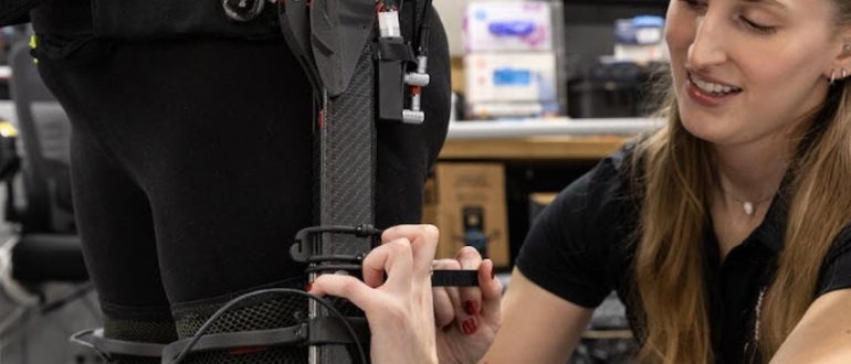
Your Stroke / Brain Injury Recovery Starts Here


ARNI home-based training and guidance for your rehab is POWERFUL. Accept no substitute.

To mark 20 years of ARNI: £50 off Full Set of Stroke Rehab 7 DVDs. Click Here to Get Yours!

News

Stroke remains a leading cause of long-term disability and a major global health burden, with current therapeutic options for acute ischemic stroke limited by a narrow therapeutic window and the formidable challenge of the blood-brain barrier (BBB). Conventional drugs often fail to penetrate the BBB effectively, resulting in poor drug bioavailability in the brain and systemic side effects.

But recent, significant preclinical and early trial data have demonstrated the potential of nanotechnology for advancing stroke treatment by overcoming key limitations of conventional therapies. Nanoparticles, designed to carry therapeutic agents across the formidable blood-brain barrier (BBB), have shown promise in delivering neuroprotective, anti-inflammatory, and thrombolytic drugs directly to ischemic brain tissue.

By encapsulating drugs, these nanocarriers can enhance stability, increase bioavailability, extend circulation time, and enable targeted delivery, thereby reducing the necessary dosage and minimizing off-target side effects. Biomimetic nanoparticles, derived from cell membranes such as platelets or neutrophils, have exhibited superior biocompatibility and targeted delivery to ischemic lesions in animal models by leveraging natural cellular homing mechanisms.

However, the successful clinical translation of this technology faces significant challenges. Further advancements are needed to improve delivery efficiency and simplify complex formulations to ensure predictable and consistent performance. Issues such as cost-effective large-scale production, inconsistent targeting, long-term stability and potential toxicity of the nanoparticles themselves must be addressed.

Bridging the gap between promising preclinical results and effective clinical therapies for future successes, requires concerted research into optimising formulations and navigating complex regulatory frameworks needed for  safe and reproducible clinical translation.

In the high-stakes world of acute stroke care, where every second counts, the speed and efficacy of intervention are paramount to saving brain cells and minimising long-term disability. A revolutionary breakthrough from Stanford University’s Department of Mechanical Engineering and Neuroimaging, published in Nature in June 2025, promises dramatic possibilities. Researchers, including Renee Shao and Jeremy Heit, have unveiled a novel spinning micro-device, the ‘milli-spinner,’ designed to remove brain clots with unprecedented precision and effectiveness.

Current thrombectomy procedures for large vessel occlusion (LVO) strokes, while effective, still have significant limitations. These methods often involve either aspiration catheters that can struggle with larger clots or stent retrievers that grapple and pull the clot, risking fragmentation and dispersal of pieces into smaller, more difficult-to-reach vessels. This can lead to incomplete recanalisation and further brain damage.

The milli-spinner, by contrast, operates on a fundamentally different and more elegant principle. As a tiny, catheter-delivered tool, it utilises a combination of localised suction, compression, and shear forces generated by its rapid spinning action to gently and efficiently process the clot.

The device works by first applying localised suction to secure the clot against the tip of the catheter. The subsequent rapid rotation then creates shear forces that cause the fibrous protein mesh of the clot (fibrin) to roll into a tight, compact ball, shrinking its volume significantly—by up to 95% in preclinical tests.

This ‘fibrin-balling’ action effectively expels the trapped red blood cells, which can then safely continue flowing, leaving behind a manageable, dense clot that is easily removed via suction. This innovative mechanism drastically reduces the risk of fragmentation and subsequent distal embolisation, which are common and serious complications of existing methods.

The results from preclinical studies using flow models and animal subjects were nothing short of remarkable. In tests on tough, fibrin-rich clots that are notoriously difficult to treat with existing devices, the milli-spinner achieved a 90% first-pass success rate in restoring blood flow, a significant leap from the 11% success rate of conventional tools for these cases.

Overall, the device more than doubled the efficacy of current technology, suggesting a major paradigm shift in interventional neurosurgery. Jeremy Heit, Chief of Neuroimaging and Neurointervention at Stanford, called the milli-spinner ‘a sea-change technology,’ projecting that it could save tens of thousands of lives and substantially reduce disability if translated successfully to clinical practice.

The potential impact of this technology extends beyond just higher survival rates. The minimally invasive and highly precise nature of the milli-spinner suggests it could also lead to better functional outcomes and a reduced risk of long-term disability, a critical factor for stroke patients and their families. By achieving faster and more complete clot removal, the device maximises the salvageable brain tissue, a key determinant of post-stroke recovery.

While the device is still undergoing further development and requires clinical trials for human use, a company has already been launched to license the technology from Stanford and accelerate its path to market. The milli-spinner represents a brilliant fusion of engineering innovation and neurovascular medicine, offering renewed hope for a faster, safer and more effective future for stroke intervention.

The recent announcement that the Queen’s Medical Centre (QMC) in Nottingham has launched a 24/7 mechanical thrombectomy service marks a significant advancement in acute stroke care and represents a critical benchmark for the UK healthcare system.

Mechanical thrombectomy, a highly effective procedure for large vessel occlusion (LVO) ischemic strokes, has revolutionised treatment by physically removing the blood clot blocking blood flow to the brain, dramatically improving functional outcomes and reducing long-term disability. However, the efficacy of this time-sensitive procedure is directly tied to its availability, and the historic limitations of service hours in many UK hospitals have meant that stroke patients presenting ‘out-of-hours’ have not always been able to access this life-changing treatment.

Nottingham’s initiative directly addresses this inequity, ensuring that every severe stroke patient in their catchment area now has the same chance at a positive outcome, regardless of when their medical emergency occurs.

The establishment of a round-the-clock service requires considerable investment in specialist infrastructure, staffing, and coordinated care pathways. It mandates a robust multidisciplinary team, including interventional neurologists or radiologists, neuroanaesthetists and specialist nurses, available at all times.

By committing these resources, Nottingham’s QMC has demonstrated a profound understanding of the “time is brain” principle. For every minute of a large vessel occlusion, millions of neurons are lost, underscoring the urgency of reperfusion. Providing 24/7 access eliminates the critical delays that previously led to preventable disability and, in some cases, death. This move aligns with and advances the recommendations of national stroke guidelines, which consistently advocate for expanded access to thrombectomy services.

While Nottingham’s accomplishment is commendable, it also highlights a persistent disparity in access to advanced stroke care across the UK. For true health equity, this model of 24/7 mechanical thrombectomy provision must be replicated in all hospitals capable of treating stroke survivors. The postcode lottery of care, where a patient’s outcome is determined by geographical location and the time of their stroke, is medically and ethically indefensible.

For example, the shocking case of Graham McGowan highlights a critical failing within Scotland’s stroke services, demanding urgent intervention by the NHS and government. Doctors carried out a brain scan which revealed a blood clot and they advised he should be treated with a thrombectomy; a procedure to remove blood clots in a large artery. But, ARI’s closest specialist thrombectomy hub, in Ninewells Hospital in Dundee, only offers the procedure from Monday to Friday, leaving Graham, a fit and active 53-year-old, with severe and preventable disability simply because his stroke occurred outside of ‘office hours’.

Tackling this failure probably requires a multi-pronged approach: immediate investment to provide a genuine 24/7 national thrombectomy service with expanded hub hours. National bodies and hospital trusts across the UK should leverage the evidence from successful centres like QMC to develop actionable strategies for commissioning and implementing their own 24/7 services.

This would not only save more lives and reduce the national burden of long-term stroke-related disability but would also align the UK’s stroke care provision with the highest international standards. The commitment shown by Nottingham is a powerful example for others to follow, demonstrating that with strategic planning and investment, equitable access to best-practice stroke treatment is an achievable goal.

Artificial Intelligence may have many downsides, but that’s certainly not the case in stroke treatment: in England, it has tripled the proportion of stroke patients who fully recover. Nearly half of stroke patients now recover to the point of functional independence, up from 16 percent.

This AI tech, which aids in the rapid analysis of brain scans, has significantly accelerated the diagnosis and treatment pathway, demonstrating how digital innovation can profoundly impact clinical outcomes. The key to this success lies in expediting the time-critical decisions necessary to administer reperfusion therapies like thrombectomy or thrombolysis.

The picture above shows on the left of each screen what 90% of doctors see using CT scans, versus the right hand side screen which shows the new technology, which identifies the problem areas automatically using the ASPECTS score card method,

One of the pivotal studies cited by NHS England involved the Brainomix e-Stroke system. This AI software was used in pilot programs across five stroke networks and has now been deployed to all 107 stroke centres in England.

Analysis of its impact showed a dramatic reduction in the time from hospital arrival to treatment, from an average of 140 minutes down to just 79 minutes. This crucial time-saving, which is critical since a stroke patient can lose millions of brain cells every minute, led to a proportional increase in positive outcomes. The proportion of patients who recovered with little or no disability soared from 16% to 48%, a threefold improvement.

The AI’s ability to provide rapid, real-time interpretation of brain scans allows specialist stroke units to make faster, more confident decisions regarding the most appropriate treatment, ultimately providing more patients with a better chance of recovering their independence.

In practice, the AI software rapidly processes CT brain scans and produces detailed reports for clinicians. These reports, including perfusion maps that highlight areas of reduced blood flow, are instantly shared across the clinical team via a secure platform. This streamlined communication and immediate insight allow for swift and coordinated action, reducing the critical time between diagnosis and the start of treatment.

The technology’s success has not only been evidenced in official reports but also validated by patient testimonials, such as that of Mr. Shawn Theoff, a retired postman from Canterbury who experienced a rapid recovery from a stroke thanks to the use of AI-enhanced diagnosis and treatment at Kent and Canterbury Hospital. He was taken to the hospital after experiencing stroke symptoms, where AI-powered decision support tools helped doctors quickly diagnose his condition and administer medication, leading to a swift recovery that saw him walking again within a few weeks. 

In-hospital strokes represent a critical and often-overlooked challenge in patient safety, with delays in detection leading to devastating outcomes. The old adage ‘time is brain’ is never more relevant than when a patient, already under medical care, suffers a new neurological event that can be easily missed amidst the complexities of a hospital environment.

This is where innovative technology like the Neuralert wristband offers a compelling and potentially game-changing solution. Recently developed at the University of Pennsylvania Health System, this wearable device continuously monitors for subtle, asymmetric arm movements (a key indicator of stroke) and automatically alerts medical staff within minutes. Unlike manual checks, which can be infrequent and prone to human error, this 24/7 automated surveillance dramatically cuts the time to diagnosis, leading to faster treatment initiation, improved patient outcomes, and reduced healthcare costs.

While the potential of Neuralert is clear, the NHS hasn’t adopted such pioneering technology. The NHS is already integrating AI for stroke detection, primarily through AI-powered software that analyses brain scans to speed up treatment decisions. These systems have, so far, proven effective in accelerating the stroke pathway from scan to treatment, but they are most impactful after a stroke has been clinically suspected and a scan requested.

Neuralert, however, addresses the crucial step before this, offering a proactive, continuous monitoring solution to bridge the gap in surveillance for high-risk, non-ambulatory patients who are difficult to monitor manually.

Despite the clear clinical and economic rationale, Neuralert hasn’t been formally planned for incorporation into NHS stroke wards, probably because the NHS adoption pathway for new medical technology is robust but slow, requiring extensive trials, regulatory approvals, and evidence of cost-effectiveness.

Therefore, while Neuralert’s utility is undeniable, its journey from a breakthrough concept to a standard feature of UK NHS stroke care will depend on successful clinical trials within the NHS, robust cost-effectiveness data, and a clear pathway for national implementation.

M (a well-known HIV drug) is an FDA-approved HIV medication, a C-C chemokine receptor type 5 (CCR5) antagonist, now being investigated for its potential to improve recovery in stroke patients. A body of preclinical research and observational studies suggests that blocking the CCR5 receptor can augment neuroplasticity, potentially enhancing functional and cognitive outcomes following a stroke. This avenue of research is particularly hopeful as it explores repurposing an existing drug with a known safety profile for a new and critical application.

Several clinical trials are currently exploring the potential of M in stroke recovery. For instance, the Canadian M Randomised Controlled Trial to Augment Rehabilitation Outcomes After Stroke (CAMAROS) is a Phase II, placebo-controlled trial evaluating the efficacy of combining M with exercise rehabilitation. The trial involves 120 participants and measures motor and cognitive function. This approach is based on animal studies showing that blocking CCR5 can enhance motor recovery and improve learning deficits after a brain injury.

Another Phase II trial focuses on preventing post-stroke cognitive impairment (PSCI) and progression to vascular dementia. Additionally, an open-label, proof-of-concept study has demonstrated the potential for M to improve post-stroke depression (PSD) symptoms.

The rationale behind these trials is compelling and grounded in basic science. A naturally occurring mutation that inactivates the CCR5 receptor (CCR5-$\Delta$32) is associated with better recovery outcomes in stroke survivors. This observation suggests that CCR5 activity can impede recovery, and therefore, blocking it with a drug like M could be therapeutically beneficial.

By inhibiting CCR5, the drug appears to promote synaptic plasticity, allowing the brain to better reorganise and repair itself after an injury. The current clinical trials, by rigorously testing these hypotheses, offer significant hope that M could one day become a valuable tool in the stroke recovery arsenal. If successful, this research could lead to the first pharmacological treatment specifically designed to enhance recovery for stroke patients..

Upper limb impairment is a common and persistent consequence of stroke, significantly affecting an individual’s independence and quality of life. The evidence shows clearly that the cornerstone of effective motor recovery is task-specific practice, a principle underpinned by evidence from neuroscience and motor learning theory. This approach posits that the brain re-organises itself in response to intensive and repetitive functional training, fostering neuroplasticity.

In the private and home rehabilitation settings, stroke survivors can engage in task-specific practice very easily by themselves or with the help of an ARNI Stroke Rehab UK trainer or physio. The rationale is that by repeatedly and intensively practicing meaningful, real-world activities, from lifting a cup to getting dressed, the brain reorganises neural pathways, improving the control over motor functions relevant to those specific tasks.

This approach moves beyond general exercises to focus directly on the functional skills that enhance independence, motivating patients through visible, goal-oriented progress. While private clinics can offer the benefit of therapist supervision, specialised equipment and intensive regimens, stroke survivors reading this will be all too aware that consistent practice at home is crucial for achieving high dosages of repetition rates necessary for effective motor learning. ARNI Stroke Rehab UK instructors work with patients to set up personalised routines, often leveraging accessible technology or adapted household items, making rehabilitation a continuous, integrated part of daily life.

The evidence base for task-specific practice is strong, and it ranks highly among recommended interventions. Moreover, national and international guidelines, such as those from the UK’s Royal College of Physicians and NICE recommend repetitive task training to improve upper limb weakness.

Correspondingly, multiple systematic reviews and meta-analyses over the years consistently report that repetitive task training yields positive, sustained improvements in mobility and upper limb function, whether implemented soon or long after a stroke. While some studies note that the effect size can be small, its efficacy often eclipses that of traditional, less focused approaches. The robust body of evidence supports task-specific practice as a cornerstone of modern stroke rehabilitation, validating its widespread use by therapists in both clinical and home environments.

For task-specific practice to be effective, it should be relevant to the survivor’s goals, performed frequently, and incorporate feedback to reinforce learning. However, traditional-type therapy has been evidenced to struggle to provide the sheer volume of high-quality repetitions needed to drive meaningful neural recovery. Correspondingly, a range of technologies have emerged fill this need to assist and optimise task-specific practice.

But stroke survivors shouldn’t view upper limb stroke rehab as a magic bullet. Tech can be an invaluable assistor, providing tools and methods to enhance and intensify these exercises, making high-repetition, focused practice more accessible and engaging. But only by using tech as an assistor to hard work, will the tech do its job. In other words task-specific training can be helped by technology but you need to get into a self rehab training programme first and understand that tech won’t work to increase function and action control after stroke without countenancing hard work.

Clinics can now days employ some very sophisticated robotic and electromechanical systems to maximise task-specific training, with many devices in the range of the average stroke survivor’s pocket. Some of these devices are robotic exoskeletons that provide adjustable arm weight support, allowing individuals with severe weakness to perform a greater range of movement. The principle of gravity compensation enables survivors to initiate and control movements themselves, rather than being passively moved, which is crucial for neuroplasticity.

For instance, ANYexo (left) is an advanced, versatile robotic exoskeleton designed for upper limb rehab in stroke survivors, enabling high-repetition training for a broad range of abilities, from survivors who are severely to those mildly affected. It features a unique kinematic structure for near-human arm movement, series elastic actuators for precise force control and intuitive programming for a wide array of daily living activities. Due to its cost it’s aimed usually at clinical settings.

For instance the Hocoma ArmeoSpring Pro (right) is a robotic exoskeleton system which provides adjustable arm weight support for the entire movement chain, from the shoulder to the hand, through a patented technology. This counterbalances gravity, allowing individuals with severe weakness to perform a greater range of movement.

The device is used in conjunction with virtual reality exercises to make therapy engaging and functional. The principle is to enable intensive, high-repetition practice of arm and hand movements in a controlled and supportive environment. The system encourages patients to initiate and control movements themselves, with the robotic arm providing just enough assistance to complete the task. This active engagement is vital for neuroplasticity.

But for stroke survivors wanting to optimise rehab at home, several other wearable and consumer-focused devices are currently available in the UK. For example, robotic gloves can offer assisted and passive training via powered mechanisms to aid grasping and releasing movements. The principle behind these home-use devices is to enable intensive, self-directed practice, leveraging biofeedback and gamification to promote plasticity. Some of these employ gamified exercises to allow users to perform high-repetition arm and hand movements, addressing the limitation of traditional therapy by increasing the dose and intensity of training outside of rehab sessions.

For instance, the Neofect Smart Glove (left), is a soft, wearable hand-and-wrist rehabilitation device that uses gamified exercises to improve motor function which incorporates sensors that track movements of the wrist and fingers, providing a platform for therapy with accompanying software offering a variety of games targeting different movements and abilities…

Another tool is a powered orthosis like the Myomo MyoPro (below) that uses electromyography (EMG) sensors to detect residual muscle signals, activating a motor to assist with arm and hand movements based on the user’s intent.

The principle here is to leverage a survivor’s own bio-signals to drive movement, creating a powerful biofeedback loop that promotes active participation and self-initiation of movement.

Like the Hocoma device, this is not a product for direct consumer purchase; rather, it’s a specialist clinical device accessed through expert rehabilitation providers. The costs for these advanced systems are substantial, reflecting their complexity and clinical application.

Functional electrical stimulation (FES) is another core technology used by therapists, sometimes integrated into other systems: FES for the upper limb delivers mild electrical stimulation to targeted muscles via a cuff, assisting with grasp and release.

The principle is to provide an external impetus for muscle contraction, which, when paired with the stroke survivor’s intent during a functional task, strengthens the neural pathways controlling movement. This helps re-educate the neuromuscular system and can enable the ability to perform task-specific practice. Clinical access to such FES systems is available through NHS and private rehabilitation services, with pricing depending on the clinical package.

An alternative, manual approach used by therapists for survivors with severe impairment is known as finger-equipped electrode electrical stimulation (FEE-ES) (left) which allows them to apply FES with precise manual control, synchronising the stimulus with even subtle, intended movement. This technique does requires a trained therapist to do it.

An example of a wearable for the home market is the Bioness H200 Wireless; (right) a sleek, wireless FES device that delivers mild electrical stimulation to specific arm and hand muscles via electrodes integrated into a soft cuff. The stimulation is controlled via an intuitive handheld unit or app, allowing for functional, task-specific training. The core principle is that FES provides an external impetus for muscle contraction, which, when paired with the patient’s intent to move, strengthens the neural pathways controlling arm and hand function.

Repetitive, intentional practice using the H200 helps re-educate muscles, reduce spasticity, and increase range of motion. In the UK, the Bioness H200 is available through private rehabilitation clinics, such as ARNI colleagues PhysioFunction and Hobbs Rehabilitation. It’s also supplied through specialist distributors like Summit Medical and Scientific. Access is typically via a clinical assessment, followed by a trial and supervised training. Pricing is substantial and depends on the specific package and clinical support required.

Others incorporating FES and EMG which are designed for survivors to purchase, like the Nura-FES, (left) are also being designed (Nura-FES is patent-pending and under investigation by long-term friend of ARNI, Professor Cherry Kilbride at Brunel University).

Unlike 25 years ago, when I had my own stroke, there is now a diverse range of technologies (from sophisticated clinical robots to consumer-friendly wearables) which can now support task-specific practice for upper limb stroke rehab. We are seeing more and more therapists (in clinical settings) able to utilise high-end robotic exoskeletons and FES systems to deliver intensive, assisted training in a controlled clinical environment, leveraging principles like gravity compensation, EMG-driven movement, biofeedback and VR to promote high-repetition re training.

Combining therapist-guided sessions with appropriate home-based wearables offers a comprehensive approach to maximising rehabilitation potential and improving functional independence. While access to high-end robotic systems remains primarily within specialist centres, more affordable wearables and digital platforms are democratising access to intensive rehabilitation, empowering survivors to take an active role in their recovery.

This burgeoning integration of wearable technology and other innovative systems within upper limb stroke rehabilitation is pretty much reshaping clinical practice and home-based recovery for UK stroke survivors. And research indicates that these technological interventions enable a higher dosage and intensity of task-specific practice, a critical driver of plasticity that has been to date limited by finite personal or clinical resources. This technological shift is not only augmenting the capacity of therapists but also offering a pathway toward more personalised, effective, and accessible rehabilitation, with the ultimate goal of improving functional independence and long-term outcomes for a larger population of stroke survivors.

For a more detailed look at new products for upper limb stroke rehab, please access ARNI Stroke Rehab UK News Channel and browse through the posts.

Nature still hides numerous ingenious solutions. DMT, or dimethyltryptamine is a natural psychoactive molecule found in many plants and mammals. According to an article published in Science Advances, DMT was found to reduce the harmful effects of stroke in animal models and cell culture experiments. The study was authored by Hungarianresearchers from the HUN-REN BRC Institute of Biophysics and Semmelweis University Heart and Vascular Centre.

DMT is also present in the human brain, and it is currently undergoing clinical trials to aid recovery of brain function after stroke. However, its exact mechanism of action had not been fully understood until now.

According to the authors of the study, the research teams found that DMT significantly reduced infarct volume and edema formation in a rat stroke model. In both animal experiments and cell culture models, DMT treatment restored the structure and function of the damaged blood-brain barrier and improved the function of astroglial cells. Furthermore, the psychoactive compound inhibited the production of inflammatory cytokines in brain endothelial cells and peripheral immune cells, while reduced the activation of brain microglia cells through Sigma-1 receptors.

The therapeutic options currently available for stroke are very limited. The dual action of DMT, protecting the blood-brain barrier while reducing brain inflammation, offers a novel, complex approach that could complement existing treatments.

Since current stroke therapies do not always result in full recovery, a DMT-based treatment may represent a promising new alternative, mainly in combination with existing methods. The recent findings from researchers in Szeged and Budapest support the development of a therapy that goes beyond the limitations of conventional stroke treatment. Clinical trials have already begun abroad, and investigation on the long-term effects of DMT are currently ongoing, but there is still a long way to go before it reaches everyday medicine.

For stroke survivors with severe upper limb paralysis, the challenge of engaging in rehabilitation is profound. Conventional electrical stimulation (ES) methods that rely on detecting residual voluntary muscle activity, such as electromyography (EMG)-triggered systems, are often unsuitable for these patients. However, a manual ES technique known as finger-equipped electrode electrical stimulation (FEE-ES) has shown promising potential. This approach allows therapists to directly and precisely control the delivery of electrical pulses, effectively reintroducing the element of patient intention into the therapeutic process.

FEE-ES is a therapist-controlled functional electrical stimulation method where the clinician wears an electrode on a finger, akin to a thimble or finger cap. This allows the therapist to manually apply and release the electrical stimulus to the patient’s skin with precise timing. By placing conventional self-adhesive electrodes on the affected limb, the therapist uses the finger-electrode to deliver the electrical pulses. This allows the therapist to precisely synchronise the electrical stimulation with the patient’s motor intention, even in cases where no voluntary muscle movement or detectable EMG signal is present. This feature engages the patient’s brain in the motor relearning process from the very beginning of rehabilitation.

Early clinical studies have shown that FEE-ES can be feasible, safe, and potentially effective for severe upper limb paresis. In a 2012 study on chronic stroke patients, those receiving FEE-ES showed greater improvement in upper extremity function compared to a control group. A  retrospective case series published this year demonstrated that FEE-ES in the acute phase of stroke was feasible, well-tolerated and associated with significant improvements in upper limb motor function in patients with severe paresis.

The synchronisation of electrical stimulation with the patient’s motor intent is believed to promote neuroplasticity, strengthening the neural pathways and encouraging long-term functional recovery. A primary advantage of FEE-ES is its ability to bridge the gap between intent and movement, a critical aspect of recovery that is often inaccessible to severely impaired stroke survivors using conventional ES systems.

FEE-ES is a clinical technique, not a standalone commercial product available for individual purchase in the UK. The implementation of FEE-ES depends on a qualified physiotherapist or occupational therapist using standard electrical stimulation equipment in a controlled, clinical setting. Therefore, its cost is integrated into the therapy sessions themselves, which vary depending on clinical setting, location, and coverage by the NHS or private insurance. Unlike an off-the-shelf device, FEE-ES is a specialised treatment modality that requires skilled professional application.

For many stroke survivors, regaining the use of their arm and hand is a significant challenge on the road to recovery. Intensive, repetitive and personalised therapy is key, but can be difficult to access consistently. Thecon Technology (HK) Limited, founded by researchers from The Hong Kong Polytechnic University (PolyU), has developed an innovative solution to this problem: the Mobilexo Arm. This wearable hybrid system combines robotics and functional electrical stimulation (FES) to accelerate and assist upper limb rehab.

The Mobilexo Arm is a portable, three-in-one rehab instrument for the paretic upper limb, designed to be used both in clinical settings and by patients at home, Unlike older FES devices with static patterns, the Mobilexo Arm uses electromyography (EMG) to detect residual electrical signals in the user’s muscles. This allows patients to control the device with their own intent, strengthening the brain’s feedback loop and encouraging neuroplasticity. It incorporates hybrid soft, inflatable components that provide mechanical assistance to the elbow, wrist and fingers. This offers a more natural and comfortable alternative to the heavy, rigid robotic arms often found in clinics.

The device provides targeted Neuromuscular Electrical Stimulation (NMES) to activate and strengthen muscles, improving coordination and sensory awareness in the affected limb. An accompanying mobile app allows patients and therapists to monitor progress remotely, track rehabilitation data in real-time, and access a variety of gamified exercises, making training more engaging and effective.

As a cutting-edge medical device, the Mobilexo Arm is not currently available for direct purchase by individuals in the UK. Thecon Technology’s current business model focuses on partnerships with hospitals and clinics. The commercialisation path for such devices typically involves extensive regulatory approvals, so its availability in the UK market would require passing these stringent tests first.

While a specific price for the UK has not been announced, the Mobilexo Arm Pro version is listed at a substantial cost in other regions. However, reports have indicated that the company is also working on a cheaper, home-based version for direct purchase by patients. For now, interested patients in the UK would need to inquire with their local stroke rehabilitation centres or explore participation in clinical trials if they become available.

The Mobilexo Arm’s main advantage lies in its hybrid design and EMG-driven control. By combining the strengths of FES and robotics into one wearable device, it offers a more sophisticated, responsive, and comfortable therapy experience.

The active participation required from the patient, guided by their own muscle signals, is far more effective at promoting neuroplasticity and functional recovery than passive, pre-set stimulation. The portability and engaging app-based exercises also make it easier for survivors to perform the frequent, high-intensity training crucial for regaining upper limb function, extending their rehabilitation beyond the clinical setting.


We are on Facebook

ARNI